
Secondary reconstruction of the orbit and conjunctival sac – a case report
Authors:
P. Drozdowski 1; A. Jaworski 2; Ł. Łątkowski 1; A. Burkacka 1; W. Lisovski 1; M. Handziak 1; K. Kott 1; K. Mildner 1; K. Brzuszkiewicz 1; A. Drozdowska 3; I. Łątkowski 1
Authors place of work:
Department of Plastic Surgery, Specialistic Medical Centre, Polanica-Zdrój, Poland
1; Department of Surgery for Child Developmental Defects and Traumatology, Faculty of Medical Sciences in Zabrze, Medical University of Silesia, Poland
2; High Medical School in Klodzko, Kłodzko, Poland
3
Published in the journal:
ACTA CHIRURGIAE PLASTICAE, 65, 1, 2023, pp. 34-36
doi:
https://doi.org/10.48095/ccachp202334
Introduction
Complete and comprehensive orbital reconstruction usually requires a two or multi-stage approach [1]. This becomes more evident in secondary reconstructions, in which previous local reconstruction options were used with poor results. In our opinion, multi-stage, reconstructive operations constitute a heavy burden for the patient, both physically and mentally. Therefore, two-stage reconstruction should be considered. We present a novel modification of vastus lateralis muscle (VLM) flap based orbital reconstitution with simultaneous implant placement for conjunctival sac reconstruction.
Case description
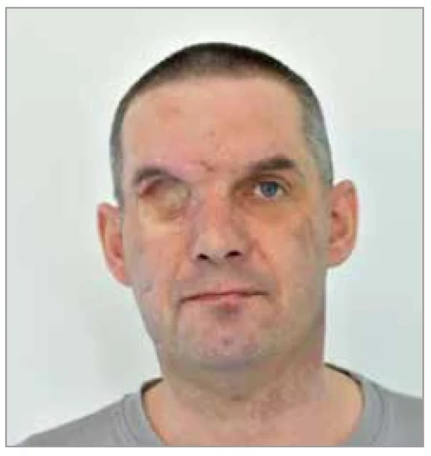
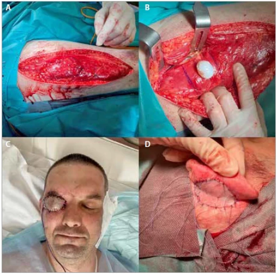
A 41-year-old patient, with a secondary defect to an injury with technical oil under high pressure, was admitted to the Plastic Surgery Department. The patient was diagnosed with a complete absence of orbital soft tissues and numerous scars after previous reconstructions with local flaps (Fig. 1), including the forehead flap. The patient underwent simultaneous reconstruction of the soft tissues of the orbit and conjunctival sac based on a prelaminated VLM flap (Fig. 2A). The flap was planned according to the ultrasionographic examination. The volume of the harvested flap was higher than the size of the defect at the recipient site to provide a robust environment around the future implant placement and because of inevitable partial muscle atrophy. Direct closure was performed at the donor site. The conjunctival sac was created utilizing an acrylic conformer implant which was wrapped with a split-thickness skin graft (STSG) from the thigh (Fig. 2B). The conformer was inserted centrally into the middle of the muscle. To create the pocket, muscle fibers were unrolled from each other, and none of them were removed. To obtain a satisfactory cosmetic effect, the flap was covered with a full-thickness skin graft (FTSG) from the retroauricular area (Fig. 2C). Due to the lack of possibility of performing direct closure of the secondary defect in the retroauricular area, it was covered with the STSG from the contralateral thigh (Fig. 2D). All of these steps were performed during one surgery. In the second operation, the eyelids were reconstructed by a simple local plasty technique (Fig. 3A–D). The postoperative course was uneventful. At present, the distant result is aesthetically and functionally pleasing (Fig. 4).


Discussion
Prelaminatied flaps are widely used in the facial plastic surgery [2,3]. Numerous techniques, including prelaminated postauricular pedicled flap, have been successfully engaged in orbital reconstruction [4]. In the case of secondary reconstructions, when previous reconstructive procedures based on local techniques and pedicle flaps have not been successful, orbital reconstruction is challenging and requires either staged, elaborated techniques [4], or microsurgery. The anterolateral thigh (ALT) flap is considered a workhorse in head and neck reconstructions due to its favorable properties, such as a large amount of skin, sufficiently long vascular pedicle, and convenient vessel diameter [5]. The disadvantages of ALT in head and neck reconstructions are the skin color mismatch [6] and the frequent need for a debulking procedure. Therefore we decided to use the modified, prelaminated ALT made up of vastus lateralis muscle wrapped around a STSG-covered mold, according to Kobus [4]. At the recipient site, we turned the flap upside down, so that the undersurface of the deep fascia faced outwards. We performed anastomoses with the superficial temporal vessels. We covered the surface of the flap with a FTSG from the retroauricular area to provide the most esthetically matching skin for the reconstructed eyelid [7]. No subsequent debulking procedure was needed [8]. The higher volume of muscle was harvested on purpose to obtain a satisfactory final volume after the future partial atrophy.
During the same procedure, we reconstructed the conjunctival sac. We placed the implant, which distinguishes this case from those described in the literature, in which it is usually reconstructed in the next stages [9]. Performing a one-stage reconstruction is associated with numerous benefits, such as lower mental and physical burden for the patient, lower costs [10], and less burden on the healthcare system. To improve the cosmetic condition, hair transplantation may be considered to reconstruct the eyelashes. We believe that future amendments to this technique will allow us to provide innveration to the flap by the connection of the motor nerve of the flap with the facial nerve to restore functionality in this area of the face.
Conclusion
To sum up, simultaneous reconstruction of the soft tissues of the orbit and the conjunctival sac is a novel technic that significantly improves the quality of life of patients after exenteration. It provokes minimal morbidity at the donor site and can also become a starting point for modifications that will allow the reconstruction of adjacent structures such as maxillary sinus, orbital innervation, or eyelashes reconstruction. At the same time, we emphasize the need to carry out more procedures to refine this method.
Conflict of interest: Authors declare no conflict of interest. All procedures performed in this study involving human participants were in accordance with the ethical standards of the institutional committee and with the 1964 Helsinki declaration and its later amendments or comparable ethical standards.
Disclosure: This case was presented at 13th Meeting of the European Plastic Surgery Research Council in Vienna on 26th August 2022.
Roles of authors
Piotr Drozdowski – conceptualization, data curation, formal analysis, investigation, methodology, project administration, supervision, writing review and editing;
Aleksander Piotr Jaworski – visualization, writing – original draft;
Łukasz Łątkowski, Karolina Brzuszkiewicz – data curation, methodology;
Krzysztof Mildner – data curation, visualization;
Aneta Drozdowska, Katarzyna Kott – data curation;
Ireneusz Łątkowski – project administration, supervision, writing review and editing;
Agnieszka Burkacka, Władysław Lisovski – visualization;
Marta Handziak – writing review and editing.
All co-authors have reviewed and approved of the manuscript prior to submission.
Aleksander Jaworski, MD
Jaworowa 5
Sosnowiec, ZIP 41 208
Silesia Voivodeship
Poland
e-mail: axjaw7@gmail.com
Submitted: 30. 8. 2022
Accepted: 19. 3. 2023
Drozdowski P, Jaworski A, Łątkowski Ł et al. Secondary reconstruction of the orbit and conjunctival sac – a case report. Acta Chir Plast 2023; 65(1): 34–36.
Zdroje
1. Sinn D.P., Bedrossian E., Vest A.K. Craniofacial implant surgery. Oral Maxillofac Surg Clin North Am. 2011, 23(2): 321–335.
2. Frias F., Horta R. Complex facial reconstruction based on 3D models: Prelamination cases and literature review. Acta Chir Plast. 2020, 62 (3–4): 86–94.
3. Dvořák Z., Cheimaris A., Knoz M., et al. Three-stage paramedian forehead flap reconstruction of the nose using the combination of composite septal pivot flap with the turbinate flap and l-septal cartilaginous graft – a case report. Acta Chir Plast. 2021, 63(1): 6–13.
4. Kobus, K. Reconstructing the eyelids after exenteration and trauma. Eur J Plast Surg. 2002, 24 : 321–327.
5. Chen Y.C., Scaglioni M.F., Carrillo Jimenez L.E., Yet al. Suprafascial anterolateral thigh flap harvest: a better way to minimize donor-site morbidity in head and neck reconstruction. Plast Reconstr Surg. 2016, 138(3): 689–698.
6. Knott P.D., Alemi S.A., Han M., et al. Skin color match in head and neck reconstructive surgery [in press]. Laryngoscope 2022, 132(9): 1753–175.
7. Wei J., Chen Q., Herrler T., et al. Supermicrosurgical reconstruction of nasal tip defects using the preauricular reversed superficial temporal artery flap. J Plast Reconstr Aesthet Surg. 2020, 73(1): 58–64.
8. Cherubino M., Berli J., Turri-Zanoni M., et al. Sandwich fascial anterolateral thigh flap in head and neck reconstruction: evolution or revolution?. Plast Reconstr Surg Glob Open. 2017, 5(1): e1197.
9. Cherubino M., Baroni T., Santoro V., et al. Social perception of reconstruction following orbital exenteration. Plast Reconstr Surg Glob Open. 2021, 9(10): e3883.
10. Lei C., Xu L., Xu F., et al. Patient satisfaction in one-stage immediate breast reconstruction after mastectomy: a multi-center comparative patient evaluation of prosthesis, LDMF, and TRAM techniques. Medicine (Baltimore) 2020, 99(22): e19991.
Štítky
Chirurgia plastická Ortopédia Popáleninová medicína TraumatológiaČlánok vyšiel v časopise
Acta chirurgiae plasticae
2023 Číslo 1
- Liečba bolesti po jednodňovej chirurgii
- Fixní kombinace tramadol/paracetamol je doporučenou volbou v léčbě chronické bolesti v ordinaci praktického lékaře
- Kombinace kodein/paracetamol prokázala stejný analgetický účinek jako hydrokodon/paracetamol
- Nová metoda kombinované analgetické léčby vychází z multimechanistické povahy bolesti
- Léčba chronické bolesti u starších pacientů vychází z farmakologických i nefarmakologických přístupů
Najčítanejšie v tomto čísle
- Pattern of invasion of oral squamous cell carcinoma and its relation to the presence of nodal metastases – a review
- Treatment algorithm for post sternotomy wound infection – our experience
- Prospective observational study of clinical outcomes in using posterior interosseous free flap for finger defects
- Role of perforator flaps in leg and foot reconstruction