
A very rare case of gastric heterotopia in the pancreas presenting as a mass
Authors:
T. Devrim 1
 ; I. Kiziloglu 2
Authors place of work:
Izmir Bakircay University, Faculty of Medicine, Department of Medical Pathology, Izmir, Turkiye
1; Izmir Private Can Hospital, Department of General Surgery, Izmir, Turkiye
2
Published in the journal:
Gastroent Hepatol 2026; 80(4): 331-335
Category:
Gastrointestinální onkologie: kazuistika
doi:
https://doi.org/10.48095/ccgh2026331
Summary
Background: Heterotopic gastric mucosa can occur anywhere along the gastrointestinal tract, from the mouth to the anus. Pancreatic GH is an exceptionally rare entity, with only limited reports available in the literature. Although it may be asymptomatic, it can also present with a variety of gastrointestinal symptoms. Case presentation: Here, we present the case of a 43-year-old woman with a suspicious cystic lesion detected on computed tomography. Histopathological examination of the distal pancreatectomy specimen revealed heterotopic gastric mucosa containing both fundic and pyloric glands, accompanied by features of chronic pancreatitis. Conclusion: By presenting this case, we aim to emphasize that heterotopic gastric mucosa, although extremely rare, can occur in the pancreas and should be considered in the differential diagnosis of pancreatic mass-forming lesions.
Keywords:
pancreas – heterotopia – gastric mucosa
Introduction
Gastric heterotopia (GH) is a rare congenital condition characterized by the presence of mature gastric mucosa in an ectopic location. Although GH may occur anywhere along the gastrointestinal tract, it is uncommon beyond the ligament of Treitz and rarely causes symptoms in adults [1,2]. GH of the intestinal tract exhibits a wide spectrum of clinicopathological presentations, often posing as a diagnostic challenge [3]. Pancreatic GH is an exceptionally rare congenital anomaly that is difficult to diagnose preoperatively [4].
GH has been reported in various regions of the gastrointestinal tract, including the tongue, oesophagus, epiglotis, small intestines, appendix, rectum, and gallbladder [3,5–8]. It is most frequently identified in the duodenum, with a reported prevalence of 1.8% in duodenal biopsy specimens [9]. GH presenting as a mass lesion has also been described in the jejunum [1,3,10], ileum [11], oesophagus [12], colon [3], and rectum [13]. In contrast, pancreatic GH is an exceptionally rare congenital malformation. By presenting this case, we aim to highlight that heterotopic gastric mucosa can also occur in the pancreas and should be considered in the differential diagnosis of pancreatic mass lesions, particularly cystic lesions.
Case report
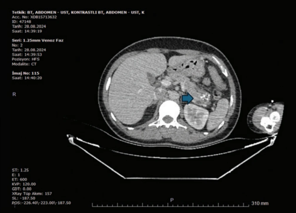
A 43-year-old woman presented with abdominal pain that developed after eating. Abdominal computed tomography (CT) revealed a 38 × 26 mm microcystic, lobulated lesion with multiple calcifications in the tail of the pancreas (Fig. 1), raising suspicion for a mucinous cystic neoplasm (MCN). Imaging findings were subsequently reviewed by a multidisciplinary team at another institution, where endoscopic ultrasonography (EUS) with biopsy was performed. Histopathological evaluation of the biopsy specimen revealed no evidence of malignancy. However, the patient subsequently presented at our institution with acute abdominal pain and guarding upon physical examination, necessitating distal pancreatectomy with splenectomy.
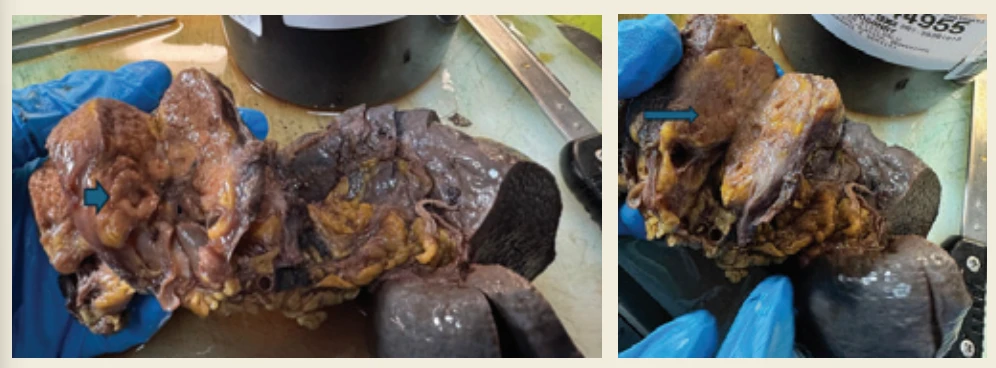
Gross examination of the pancreatectomy specimen revealed a well-demarcated yellow-pink lesion measuring 5 cm in the widest dimension, separated from the surrounding pancreatic parenchyma by a relatively regular border. Within the central portion of the lesion, a 1.5 cm raised area with a distinct gross appearance was identified. Adjacent to this, a 1 cm cyst with a smooth inner surface was observed near the serosal aspect. Multiple additional millimetric cystic structures were present throughout the remaining lesion (Fig. 2).
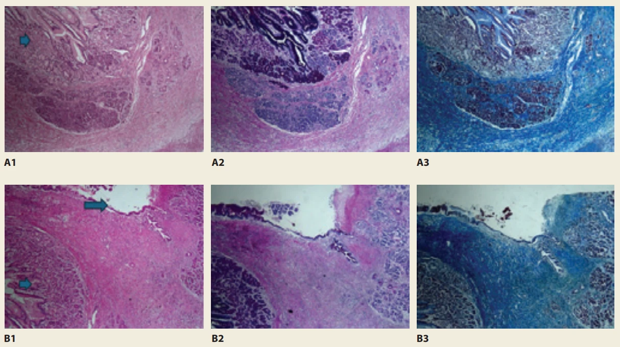
Microscopic examination revealed a 1.5 cm focus of gastric heterotopia within the pancreatic parenchyma. In the adjacent lesion, the pancreatic tissue showed fibrosis and mild-to-moderate inflammatory cell infiltration composed predominantly of scattered lymphocytes, with fewer eosinophils and plasma cells. In addition, pseudocyst formation, ductal dilatation with focal destruction, and proteinaceous material with focal calcification were observed, consistent with features of chronic pancreatitis (Fig. 3).
Discussion
GH is generally considered a congenital developmental anomaly characterized by the presence of ectopic gastric mucosa. Histologically, it comprises the full thickness of the gastric mucosa, including specialized fundic glands containing chief and parietal cells, with the surface lined by gastric foveolar epithelium [3]. GH has been reported in various locations, including the oral cavity, oesophagus, duodenum, jejunum, ileum, rectum, anal canal, gallbladder, and bile duct. It occurs most frequently in the first and second portions of the duodenum [2,3,5–8,10,11,13].
GH is generally considered a congenital developmental anomaly, although its precise aetiology remains uncertain. The most widely accepted hypothesis is that it results from abnormal differentiation of pluripotent primitive endodermal stem cells during embryogenesis [14]. During normal development, the primitive gut, comprising of the foregut, midgut, and hindgut, arises from the endoderm of the gastrula. Both the stomach and pancreas originate from the distal foregut [4]. In the oesophagus, heterotopic gastric mucosa is thought to result from incomplete replacement of the primitive columnar epithelium by stratified squamous epithelium during embryonic development, leading to the persistence of gastric mucosa within the oesophageal wall [14]. Similarly, aberrant differentiation of primitive endodermal cells may result in ectopic gastric mucosa at various sites throughout the gastrointestinal tract. Although ectopic pancreatic tissue within the stomach is relatively well recognized, the presence of heterotopic gastric mucosa within the pancreas is exceptionally rare [4].
Heterotopic lesions are difficult to diagnose accurately using imaging modalities or endoscopic visualization because they lack distinctive features [3]. Classification of pancreatic cystic lesions remains challenging. Although CT and magnetic resonance imaging (MRI) are effective for accurately localizing these lesions, their ability to characterize them in detail is limited. EUS plays a crucial role in the evaluation of pancreatic cystic lesions by providing high-resolution imaging of the cyst wall, internal septations, mural nodules, and their relationship to the pancreatic ductal system [4]. Nevertheless, a definitive diagnosis often requires histopathological examination. Surgical excision of heterotopic lesions generally results in complete symptom resolution and is considered curative in most cases. Consequently, histopathological examination remains the gold standard for diagnosis, and long-term follow-up is generally unnecessary after complete excision [3].
Pancreatic cystic lesions are frequently detected incidentally during CT or MRI performed for various indications. Accurate characterization is essential, as clinical management depends on differentiating neoplastic from non-neoplastic cysts. Although CT and MRI are effective for lesion detection, EUS provides superior morphological assessment. Pancreatic cystic lesions comprise a heterogeneous group of entities, including pseudocysts and neoplastic cysts such as MCNs, intraductal papillary mucinous neoplasms, serous cystic neoplasms, solid pseudopapillary neoplasms, and cystic pancreatic neuroendocrine tumours. Because these lesions often exhibit overlapping imaging features, accurate differentiation may require EUS combined with cyst fluid biochemical, cytological, and molecular analyses. Recent advances, including next-generation sequencing (NGS) and artificial intelligence (AI) -assisted image analysis, have further enhanced the diag-nostic evaluation of pancreatic cystic lesions [15–17].
In the present case, EUS contributed to the evaluation of the pancreatic cystic lesion, although definitive diagnosis of GH was established by histopathological examination following surgery. Consistent with the findings of Chen et al. (2020), EUS may play an important role in the assessment of benign pancreatic cystic lesions and rare congenital anomalies such as GH. To date, pancreatic GH has been reported only rarely [4]. Our study adds to the limited evidence indicating that GH can occur in unexpected locations. Clinicians should consider this rare entity in the differential diagnosis of unusual pancreatic cystic lesions, particularly when common conditions such as MCN are suspected.
Conclusion
The present case highlights the importance of including GH in the differential diagnosis of pancreatic lesions, particularly those with cystic features. Recognizing this rare entity in unusual anatomical locations may help improve diagnostic accuracy and guide appropriate clinical management.
Submitted/Doručeno: 21. 5. 2026
Accepted/Přijato: 6. 8. 2026
Corresponding author
Assoc. Prof. Tuba Devrim, MD
Izmir Bakircay University
Cigli Education and Research Hospital
Yeni Mahalle, 8780/1
Sk. No: 18
35620 Cigli/Izmir, Turkiye
tuba.devrim@bakircay.edu.tr
Zdroje
1. Anderson J, Facey M. Gastric heterotopia as the cause of distal small bowel obstruction: a case report. Pathology 2024; 56 : 45.
2. Lee W, Jee YS. Jejunal gastric heterotopia presenting as perforation peritonitis in a middle-aged adult: A case report. Int J Surg Case Rep 2021; 80 : 105625. doi: 10.1016/j.ijscr.2021.02.011.
3. Nasir A, Amateau SK, Khan S et al. The many faces of intestinal tract gastric heterotopia; a series of four cases highlighting clinical and pathological heterogeneity. Hum Pathol 2018; 74 : 183–187. doi: 10.1016/j.humpath.2018.01.003.
4. Chen Q, Chai N, Linghu E. A rare case of the pancreas with heterotopic gastric mucosa detected by EUS (with video). Endosc Ultrasound 2020; 9 (6): 410–412. doi: 10.4103/eus.eus_19_19.
5. Estes K, Dhall D, Weber F. An Unusual Duodenal Finding. Gastroenterology 2023; 164 (2): 194–195. doi: 10.1053/j.gastro.2022.07.074.
6. Khan S, Niaz S, Khan M et al. A Case of Heterotopic Gastric Tissue in Duodenal Bulb: An Interesting Endoscopic Finding. Cureus 2022; 14 (4): e24271. doi: 10.7759/cureus.24271.
7. Maione F, Chini A, Gennarelli N et al. Endoscopic resection of a large polypoid gastric heterotopia of duodenum: a case report. Clin Case Rep 2021; 9 (8): e04633. doi: 10.1002/ccr3.4633.
8. Yeldir N, Bozbıyık N, Özer H et al. Gastric heterotopia in the gallbladder: Mimicking the tumor imagine in radiology. CMJ 2021; 43 (4): 427–430.
9. Genta RM, Kinsey RS, Singhal A et al. Gastric foveolar metaplasia and gastric heterotopia in the duodenum: no evidence of an etiologic role for Helicobacter pylori. Human Pathology 2010; 41 (11): 1593–1600. doi: 10.1016/j.humpath. 2010.04.010.
10. Bhattacharya B, Jakate S, Saclarides TJ et al. Gastric heterotopia presenting as a mass in jejunum. Arch Pathol Lab Med 2003; 127 (4): 506–507. doi: 10.5858/2003-127-0506-GHPAAM.
11. Carlson Q, Golconda U, Sun B. Gastric heterotopia with perforation mimicking neoplastic process in ileum. J Surg Case Rep 2020; 2020 (7): rjaa224. doi: 10.1093/jscr/rjaa224.
12. Ward EM, Achem SR. Gastric heterotopia in the proximal esophagus complicated by stricture. Gastrointestinal Endoscopy 2003; 57 (1): 131–133. doi: 10.1067/mge.2003.44.
13. Dinarvand P, Vareedayah AA, Phillips NJ et al. Gastric heterotopia in rectum: A literature review and its diagnostic pitfall. SAGE Open Med Case Rep 2017; 5 : 2050313X17693968. doi: 10.177/2050313X17693968.
14. Yang G, Ren H, Xiong T et al. Simultaneous heterotopia of the gastric mucosa and pancreatic tissue leads to perforation of the small intestine: case report and literature review. AME Case Rep 2026; 10 (41): 22. doi: 10.21037/acr-24-236.
15. Singh S, Chandan S, Vinayek R et al. Endoscopic techniques for the diagnosis of pancreatic cystic lesions. World J Gastroenterology 2025; 31 (1): 101082. doi: 10.3748/wjg.v31.i1.101082.
16. Guilabert L, Nikolìc S, de-Madaria E et al. Endoscopic ultrasound for pancreatic cystic lesions: a narrative review. BMJ Open Gastroenterology 2025; 12 (1): e001893. doi: 10.1136/bmjgast-2025-001893.
17. Liu E, Yang F. EUS-Anchored Multimodal Evaluation of Pancreatic Cystic Lesions: Toward a Conceptual Diagnostic Framework. J Clin Med 2026; 15 (10): 3893. doi: 10.3390/jcm15103893.
Štítky
Detská gastroenterológia Gastroenterológia a hepatológia Chirurgia všeobecnáČlánok vyšiel v časopise
Gastroenterologie a hepatologie
2026 Číslo 4
- Těhotenství a idiopatické střevní záněty – terapie zlepšuje stav a je bezpečná
- Vliv těhotenství na klinickou aktivitu Crohnovy nemoci
- Medikace u IBD v těhotenství
- Adherence a kvalita života adolescentních pacientů s IBD
- Adherence pacientů se střevním zánětlivým onemocněním
Najčítanejšie v tomto čísle
- Selecting the right probiotic: from strain specificity to rational clinical application
- Klinická a experimentální gastroenterologie
- Severe upadacitinib-induced anemia in a multi-refractory Crohn’s disease patient – a case report
- Farmakologická léčba obezity u kandidátů transplantace jater: první zkušenosti