
Study of KDM1A and VEGF changes as the responsible genes in the angiogenesis of breast cancer
Studie změn KDM1A a VEGF jakožto genů zodpovědných za angiogenezi karcinom prsu
Východiska: Nejnovější poznatky ohledně podílu mikroRNA na nádorové angiogenezi a onkogenních účincích mikroRNA ukazují na jejich případnou roli v angiogenezi karcinomu prsu. Exozomy získané z nádoru jsou považovány za bohatý zdroj mikroRNA, které dokáží regulovat ostatní buňky v nádorovém mikroprostředí, vč. vaskulárních endoteliálních buněk. Tato studie analyzuje účinek chemoterapie tamoxifenem na expresi klíčové microRNA, miR-329, a upozorňuje na spojitost mezi touto mikroRNA a genem KDM1A, který se účastní dráhy vaskulárního endoteliálního růstového faktoru (VEGF).
Materiál a metody: Byly zakoupeny buňky karcinomu prsu MCF-7 a kultivovány v kompletním živném médiu. K těmto buňkám byl přidán tamoxifen a pak byly z média extrahovány jejich exozomy. Z nich byly izolovány RNA a pomocí metody polymerázové řetězové reakce (polymerase chain reaction – PCR) v reálném čase byla zkoumána exprese genů miR-329, VEGF a KDM1A v exozomech.
Výsledky: Výsledky této studie ukázaly, že při léčbě tamoxifenem se v exozomech získaných z buněk karcinomu prsu MCF-7 zvýšila exprese genů miR-329. Exprese genů KDM1A a VEGF v exozomech buněk ošetřených léčivem byla downregulovaná.
Závěr: Výsledky tohoto experimentu ukázaly, že po přidání tamoxifenu k buňkám karcinomu prsu dochází vlivem zvýšení miR-329 ke snížení exprese VEGF a KDM1A. Tím se snižuje angiogeneze a uplatňují se tak protinádorové účinky tohoto léku.
Klíčová slova:
karcinom prsu – nádorová angiogeneze – exozom – miR-329 – tamoxifen
Authors:
N. Hashemi; M. Kavousi; F. Jamshidian
Authors place of work:
Department of Biology, East Tehran Branch, Islamic Azad University, Tehran, Iran
Published in the journal:
Klin Onkol 2023; 36(4): 281-286
Category:
Původní práce
Summary
Background: Recent developments regarding the contribution of microRNAs to tumor angiogenesis and the oncogenic effects of microRNAs point to their potential role in breast cancer angiogenesis. Tumor-derived exosomes are considered a rich source of microRNAs that can regulate the function of other cells in the tumor microenvironment, including vascular endothelial cells. This study analyzes the effect of tamoxifen chemotherapy on the expression of a key microRNA, miR-329, and introduces a regulatory link between this microRNA and the KDM1A gene associated with the vascular endothelial growth factor (VEGF) messaging pathway.
Materials and methods: MCF-7 breast cancer cells were purchased and cultured in a complete culture medium. These cells were treated with tamoxifen and then their exosomes were extracted from the culture medium. The RNAs of the exosomes were isolated and the expression of miR-329, VEGF, and KDM1A genes in the exosomes was investigated using the real-time polymerase chain reaction (PCR) method.
Results: The results of this study showed that tamoxifen treatment increased the expression of miR-329 in exosomes derived from MCF-7 cancer cells. The expression of KDM1A and VEGF genes in drug-treated cell exosomes is downregulated.
Conclusion: The results of this experiment demonstrated that the treatment of breast cancer cells with tamoxifen reduces the expression of VEGF and KDM1A by increasing miR-329. The treatment therefore reduces angiogenesis, and thus its anti-tumor effects are applied.
Introduction
Breast cancer occurs as a result of the abnormal growth of cells in the breast. That growth pattern stops at a certain stage in benign tumors, whereas the malignancy development continues uncontrollably in malignant tumors [1].
Tamoxifen is a drug used to reduce or stop the effects of the female hormone, estrogen, in the body and is one of the drugs used in breast cancer treatment [2].
Exosomes are small vesicles (30–100 nm) that can transfer lipids, proteins, mRNAs, and microRNAs to the receptor cell when moving to other cells. Cancer cells have been shown to secrete far more exosomes than normal cells, thus transmitting tumor-dependent signaling molecules to surrounding cells [3].
MicroRNAs are a class of endogenous non-coding RNA molecules that regulate gene expression at the post-transcriptional level by mRNA degradation and/or translation inhibition. There exist more than 2,000 miRNA encoding genes in the human genome, which make up about one percent of the whole genome [4]. MicroRNAs are among the molecules being transferred by exosomes and regulate most of the cell‘s biological functions. They regulate translation and transcription programs and coordinate pathological as well as physiological processes such as development, cell differentiation, proliferation, apoptosis, and tumor growth [5]. MicroRNAs act as small guide molecules in RNA silencing and regulate the expression of various genes, both at the mRNA level and the protein level, through degrading the target mRNA and silencing the translation. One of the recent developments in this field of study is the understanding of their role in oncogenesis. Changes in the expression and regulation level of microRNAs play a role in the initiation, progression, and metastasis of various tumors. Biochemical and genetic evidence confirms the role of microRNAs in tumor development. The abnormal expression and regulation of mRNAs in cancer cells is a well-known phenomenon, the cause of which is yet unidentified [6].
The exact mechanism by which tumor cell-derived exosomes affect their constituent microenvironmental cells, including endothelial cells, has not yet been determined, and the biological function of exosomal microRNAs in receptor cells is not well understood. Therefore, this study investigates the effect of tamoxifen chemotherapy drug on the expression of a key microRNA involved in angiogenesis, miR-329, in tumor exosomes and introduces a regulatory relationship between this microRNA and KDM1A gene, associated with VEGF messaging pathway.
Materials and methods
Cell preparation and culture
The MCF-7 breast tumor cell-line with NCBI CODE 135 and human umbilical vein endothelial cells (HUVEC) cell-line were both purchased from the Iranian Genetic Resources Center. Subsequently, the cells were cultured in full culture medium containing fetal bovine serum and trypsin (GibCo). The flasks were kept inside an incubator with CO2 saturation rate 5% and a temperature of 37 °C.

Tamoxifen treatment
In order to reach the appropriate dosage of tamoxifen, cell viability was evaluated by the half maximal inhibitory concentration (IC50) through applying 3 - (4,5dimethylthiazole-2-yl) -2,5-diphenyltetrazolium bromide (MTT) to MCF-7 cell line. In brief, almost 10,000 cells/well were seeded into a 96-well plate in RPMI-1640 medium and were allowed to attach overnight. To increase the accuracy of the operation, 3 wells were dedicated to each condition. Subsequently, the cells were treated with different ascending doses of tamoxifen and incubated for 24, 48, and 72 h (37 °C, 5% carbon dioxide in air). Cell growth inhibition was determined using the MTT assay. The medium was removed and 20 µL of MTT (5 mg/mL) (Sigma, St. Louis, MO, USA) was added to each well of 96-well plate and were incubated for 4 h at 37 °C. Composed crystals dissolved by adding 200 µl of dimethyl sulfoxide (DMSO) to each well. Finally, the optimal absorbance level was measured by the ELISA-reader machine at 570 nm. The MTT assays were performed at least three times for each concentration of drug and the percentage of surviving cells relative to control (untreated sample) was determined. According to MTT assay, the IC50 of tamoxifen after 48 h was determined as 54 μM. Tamoxifen was dissolved in DMSO and then added to culture medium to the final desired concentration based on the determined IC50 and filtered. The cells (at 80% confluency) were incubated with freshly prepared drugs for 48h in a humidified incubator before being trypsinized and washed with phosphate-buffered saline three times and stored at −70 °C.
Exosome isolation
Exosomes were isolated from the culture medium of MCF-7 cells. For this purpose, the culture medium was first centrifuged at 3,000 g for 10 min to separate the dead cells from the culture medium. Exoquick solution (System Biosciences, CA, USA) was then added to the supernatant. Vortex binds the exoquick solution to exosomes. Then it was incubated for 20 min at 4 °C. The centrifugation was afterwards performed at a speed of 1,500 g for 15 min. 50 μL phosphate buffered saline (PBS) was then added to the solution and pipetting was performed. The content was transferred to a microtube and placed on a heater for 5 min at 70 °C and then at −20 °C. Exosome isolation was performed from the supernatants of both treated and control cell groups, confirmation of which was done by microscopic examination, digital electron microscope (Digital FESEM, KYKY-EM3200, China), evaluating the size and morphology of the exosomes.
Exosome labeling
PKH26 (PKH26 Red fluorescent – Mini26-1, Sigma Aldrich, USA) was used to label the exosomes. 1 mL of the exosomes was transferred to a microtube, and 1 mL of Diluent C was added to it and gently pipetted. From the diluted paint PKH26, 1 mL was added into the microtube containing the exosome and then incubated at room temperature for 5 min. To stop the labeling, 2 mL of Diluent C was again added into the microtube and placed at room temperature for 5 more min. Then the exoquick solution was added with a ratio of 3 : 1 and kept at 4 °C for 20 min. Later, the solution was centrifuged for 20 min at 15,000 g. After centrifugation, the top solution was removed and the exosome-containing precipitate was dissolved in 30 μB PBS. The endothelial cells were then treated with stained exosomes, followed by incubation at room temperature for 12 h. Due to the presence of miRNAs in exosomes, the exosomal transmission of miR-329 to endothelial cells was investigated. In this regard, endothelial cells were treated by 100 µg/mL of tumor exosomes and 50 µg/mL of alpha-amanitin. The rate of miR-329 transferring to endothelial cells was evaluated at 0, 24, and 48 h post-treatment. The reason for using alpha-amanitin is to inhibit any transcription activity that can be induced by exosomal treatment in cells.
Exosome authenticity
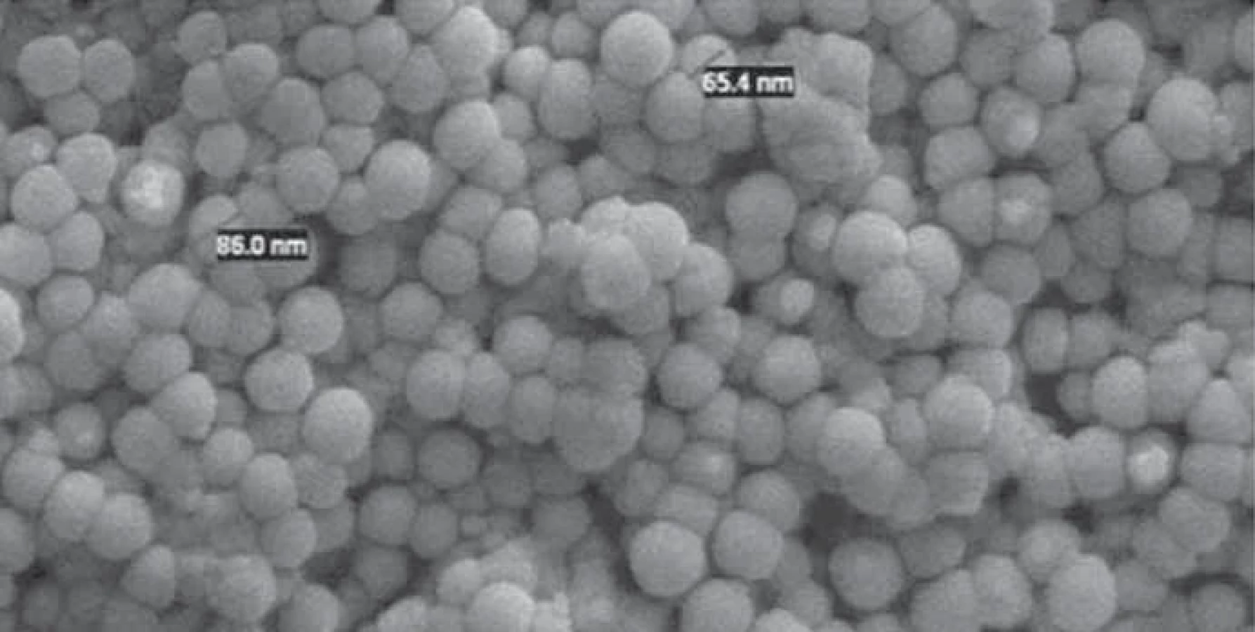
The morphology and size of the purified exosomes were measured by scanning electron microscopy (Digital FESEM, KYKY-EM3200, China). For this purpose, a fraction of above-mentioned exosomes was fixed in 2.5% glutaraldehyde on a microscope slide, washed by PBS, and an ascending amount of ethanol was utilized for critical point dehydration. The slide was then dried on a glass substrate and coated with a thin layer of gold.
RNA isolation
RNA isolation from cells and exosomes was performed using the Trizol (Invitrogen) agent. After centrifuging the cells and adding 50 μL of Trizol solution, 15 μL of chloroform was added to it and incubated at room temperature for 2–3 min. After centrifugation at 12,000 g, the upper phase was removed and isopropanol was added and then the whole solution was incubated on ice for 10 min. The centrifugation was performed another time at 12,000 g for 10 min. Subsequently, 1 mL of 75% ethanol was added to the residual precipitate, and centrifugation was performed again at 7,500 g at 4 °C for 5 min. The RNA sample was stored in a freezer at −80 °C. The quantitative analysis of the isolated RNA was done using spectrophotometry (NanoDrop Technologies, Wilmington, DE, USA).
Investigating the expression level of KDM1A, VEGF, and miR-329
cDNA was generated using the first strand cDNA Synthesis kit (Thermo Fisher Scientific, MA, USA), Oligo dT, and random hexamer primers. In order to perform the real-time polymerase chain reaction (PCR), the forward and reverse primers (Tab. 1), cDNA, DEPC water, Mastermix, and CybrGreen dye were all added and the solution was placed in a thermocycler. Data analysis was performed using the threshold cycle comparison method according to the table below. Normalization of changes in miRNA expression level was done in comparison with the endogenous U6 snRNA expression level, while the expression level of other genes was compared with glyceraldehyde-3-phosphate dehydrogenase (GAPDH). Data analysis was performed using ABI step one Real-Time PCR Software v2.0.2 (Applied Biosystems, UK) software, Excel software (Microsoft, Redmond, WA, USA, 2010), and GraphPad software.
Statistical analysis
Statistical analysis of the obtained data was performed using SPSS v.20 software (IBM, Armonk, NY, USA) and Anova test and Tukey follow-up test. P < 0.05 was considered significantly meaningful in this study.
Results
Exosome isolation confirmation
The size and morphology of the exosomes were evaluated by scanning electron microscope (SEM). The results showed that the isolated exosomes had a spherical appearance with a size range of 30–150 nm (Fig. 1).

Measurement of endothelial cells‘ ability to absorb PKH26 fluorescent - -labeled tumor exosomes
In order to determine the ability of endothelial cells to absorb tumor exosomes, HUVEC endothelial cells were treated by PKH26-labeled fluorescent colored exosomes. They were then incubated for 12 h. Fluorescent microscopic analysis showed that PKH26 fluorescent-labeled exosomes are located in the cytoplasm of endothelial cells (Fig. 2). This indicates the ability of HUVECs to absorb endosomes derived from breast tumor cells.
Examination of miR-329 expression level in exosomes isolated from MCF-7 breast cancer cells
To investigate the angiogenic effects of exo-MCF-7 on endothelial cells, we evaluated KDM1A and VEGF mRNA expression levels by RT-qPCR, relative to GAPDH as the housekeeping gene, in HUVECs following treatment with exo-MCF-7 (100 µg/mL) or vehicle control (PBS). This study showed that exo-MCF-7 can upregulate transcript levels of KDM1A and VEGF in HUVECs in a time-dependent manner (Graph 1). Consistently, exo-MCF-7 had also a significant influence on VEGF protein levels in HUVECs (Graph 2). Moreover, this study also revealed an increase in VEGF protein secretion of endothelial cells treated with 100 µg/mL of exosomes after 48 h. Thus, the increased VEGF protein expression and secretion after exo-MCF-7 treatment was consistent with its augmented mRNA expression levels. According to Graph 2, the miR-329 expression level in tumor cell-derived exosomes treated with tamoxifen has a higher expression than exosomes derived from the control cell group. This result suggests that tamoxifen treatment may alter the expression profile of miRNAs which have tumor suppressor activities such as miR-329 in tumor cell-derived exosomes. The expression level of miR-329 being treated with tamoxifen has upregulation. All experiments were conducted three times independently.
Transferring miR-329 from MCF-7 breast tumor cell-line to HUVEC endothelial cells
Initially, the exosomal transfer of miR-329 to endothelial cells was investigated due to the presence of miRNAs in the exosomes. For this reason, HUVEC endothelial cells were first treated by 100 µg/mL alpha-amanitin, and then the rate of miR-329 transfer to endothelial cells was assessed at 0, 24, and 48 h post-treatment. The reason lying behind using alpha-amanitin is to inhibit any transcription activity that can be induced by exogenous exosomes in the endothelial cells. As it is seen in Graph 1, the level of miR-329 transcripts in endothelial cells treated by exosomes and alpha-amanitin increased significantly, more than twice, over time.
Effects of breast tumor cells-derived exosomes on the expression level of KDM1A and VEGF genes
Tumor-derived exosomes that have been left untreated induced the expression level of KDM1A and VEGF genes time-dependently. Much to our surprise, in contrast, tamoxifen-treated tumor exosomes reduced KDM1A and VEGF expression levels over time. Therefore, given the greater presence of miR-329 in tamoxifen-treated tumor exosomes, it is comprehended that the miR-329 exosomal transfer reduces the expression level of KDM1A and VEGF in endothelial cells after 24 and 48 h significantly and time-dependently. Therefore, it is assumed that the exosomal transfer of miR-329, which is a tumor suppressor, from the mammary tumor cells being treated by tamoxifen may have an inhibitory effect on angiogenesis through decreasing the activity and expression level of KDM1A and VEGF pro-angiogenic genes (Graphs 5, 6).
Discussion
Cancer cells seek to activate regulatory pathways for survival and development, as well as to obtain the metabolites and growth factors they need [7]. Since angiogenesis is developed by microenvironmental changes in cancer cells through tumor exosomes, it appears that inhibiting angiogenesis is mediated by the anti-angiogenic chemotherapy drug, tamoxifen, through modulating related miRNAs‘ expression. Therefore, it is assumed that tumor cell exosomes should be targeted to inhibit angiogenesis [8]. Tumor-derived exosomes are considered as a rich source of miRNAs that could regulate the function of other cells in the tumor microenvironment, including vascular endothelial cells. However, the exact mechanisms by which tumor cell-derived exosomes affect their microenvironmental cells, as well as the biological function of exosomal miRNAs in cells, have not been well established [9]. Therefore, this study evaluated the effect of one of the chemotherapy drugs, tamoxifen, on the expression level of a key microRNA involved in angiogenesis, miR-329, and tried to introduce a regulatory relationship between this microRNA and KDM1A gene associated with the VEGF messaging pathway. The results showed that the expression of miR-329 in the exosomes derived cells treated by tamoxifen increased significantly and time-dependently. On the other hand, the expression of KDM1A and VEGF genes in drug-treated cell exosomes had a downward pattern.
In 2015, Tahakashi et al. investigated the breast tumor-derived miRNAs and introduced these microRNAs as a diagnostic biomarker in the treatment of breast cancer [10]. The result of the present study, in line with the research of Tahakashi et al., confirm the role of miR-329 as a diagnostic biomarker in breast cancer. In 2015, Hannafon et al. concluded that the use of the anti-cancer drug docosahexaenoic acid (DHA) changes the exosomal secretions of breast cancer cells. They isolated MCF7 and MDA-MB-231 breast cancer cell exosomes after treatment with DHA and observed an increase in the secretion of exosomes and the content of miRNAs of DHA-treated exosomes. The expression level of miRNA-83 in MCF7-treated exosomes had changed. The group concluded that DHA alters the exosomal secretions of breast cancer cells and their miRNA content, thereby inhibiting angiogenesis [11]. Although the chemotherapy drug used in the present study, tamoxifen, is different from that of Hannafon et al., the results are in line with each other in terms of diminishing the expression level of genes targeting the angiogenesis and cancer development.
Recent research suggests that KDM1A is an endothelial biomarker that promotes angiogenesis. It has recently been discovered that KDM1A is a transferring inducer between the epithelium and the mesenchymal tissue, thus expanding the invasion of breast cancer cells. Tumor secretions stimulate the activation of p38 MAPKs / NF-kB, and activation of many gene-producing genes including VEGF, MMP-9, and IL-8. AA98, which is the anti-molecular antibody, interferes with this signaling. The association of KDM1A with tyrosine kinase P59, phosphorylate P125, and paxillin promotes the migration of endothelial cells. KDM1A promotes angiogenesis in both laboratory and somatic cell conditions [12]. Increasing the expression level of KDM1A facilitates the endothelial response to the kinase Src stimulated by VEGF or VEGF-induced Src kinase family (SKF), p38 mitogen-activated protein kinase (MAPKs) and NF-kappaB activation, and subsequently increases the migration of endothelial cells and tube formation. CD146 is a co-receptor for VEGFR-2; therefore, suppression of CD146 by miR-329 impairs signaling and angiogenesis induced by VEGF [13].
The expression level of miR-329 decreases in various tumors and its re-expression in the cell prevents the growth and development of melanoma. A characteristic of rheumatoid arthritis, which is a chronic autoimmune disease, is angiogenesis and cartilage decay, in which miR-329 has a role [14]. miR-578 and miR-329 play a role in the process of angiogenesis in breast cancer associated with the BRCA gene. Laboratory evaluations on HEK293, MCF-7, and SUM149PT cells to investigate the role of miR-578 and miR-329 have shown that these miRNAs have been suppressed in breast cancer associated with BRCA 1 & 2. miR-578 and miR-329 are associated with the central connection, or focal adhesion, the VEGF vascular endothelial growth factor, and the signaling pathway of hypoxia inducible factor-1 (HIF-1). miR-578 and miR-329 control angiogenesis in breast cancer associated with BRCA 1/2 and do so by regulating the vascular endothelial vascular growth factor VEGF and factor 1 by inducing HIF-1 [15]. In line with this research, the present study showed that when the tamoxifen-treated exosomes were transferred to the endothelial cell line, the expression of VEGF gene had a decreasing pattern.
Based on the results of the present study on the MCF-7 breast cancer cell line, it was found that there was a significant relationship between 54 μM dose of tamoxifen and MCF7 cell supernatant exosomes, in a way that the expression level of exosomal miR-329 showed a significant increase. In this study, the effect of tamoxifen-treated tumor-derived exosomes was evaluated on the HUVEC endothelial cell line to assess the process of angiogenesis and treatment of breast cancer in its early stages. Applying the experiment group to the endothelial cell line reduces the expression of KDM1A gene in endothelial cells significantly over time. The second gene studied in this study was VEGF, the result of which was the same as KDM1A. Ultimately, this paper could conclude that the exosomal transfer of miR-329 to endothelial cells reduces the expression of genes responsible for the angiogenesis process, including VEGF and KDM1A, identification of which could mitigate the tumor process.
When breast cancer cells are treated with tamoxifen, the level of miR-329, which is a tumor suppressor microRNA in the exosomes of these cells, is much higher than in untreated cancer cells. In contrast, when tamoxifen-treated tumor-derived exosomes are transferred to endothelial cells, the expression level of KDM1A and VEGF genes declines in a time-dependent manner. This achievement brings us to the idea that that the therapeutic effect of tamoxifen on breast cancer lies behind its relationship with the expression level of exosomal miRNAs that regulate the cancer microenvironment.
Conclusion
The results of this experiment demonstrated that the treatment of breast cancer cells with tamoxifen, by increasing miR-329, reduces the expression of VEGF and KDM1A and thus reduces angiogenesis, and thus its anti-tumor effects are applied.
Acknowledgments
This study was conducted at the Genetic, cellular and molecular laboratory of Islamic Azad University - East Tehran Branch (Ghiamdasht), thus appreciating the spiritual support of this center is admitted.
Availability of data and materials
The data used in this study are available and could be demanded from the corresponding author.
Competing interests
The authors declare that they have no competing interests.
Authors’ contributions
Niloufar Hashemi: conceptualization, methodology, software, validation, data curation, writing – review and editing;
Dr. Mahsa Kavosi: validation, formal analysis, conceptualization, writing – original draft, investigation, methodology, resources;
Dr. Faranak Jamshidian: conceptualization, supervision, project administration, methodology, investigation, data curation, visualization, writing – original draft, validation, formal analysis, conceptualization, resources, data curation, software.
Consent for publication
All the authors confirm that the manuscript represents their honest work.
Zdroje
1. Osborne CK. Tamoxifen in the treatment of breast cancer. N Engl J Med 1998; 339 (22): 1609–1618. doi: 10.1056/NEJM199811263392207.
2. Folkman J. Fundamental concepts of the angiogenesis process. Curr Mol Med 2003; 3 (7): 643–651. doi: 10.2174/1566524033479465.
3. Cristofanilli M, Charnsangavej C, Hortobagyi GN. Angiogenesis modulation in cancer research: novel clinical approaches. Nat Rev Drug Discov 2012; 1 (6): 415–426. doi: 10.1038/nrd819.
4. Folkman J. Angiogenesis. Annu Rev Med 2016; 57 : 1–18. doi: 10.1146/annurev.med.57.121304.131306.
5. Makrilia N, Lappa T, Xyla V et al. The role of angiogenesis in solid tumors: an overview. Eur J Intern Med 2019; 20 (7): 663–671. doi: 10.1016/j.ejim.2009.07.009.
6. Bergers G, Benjamin L. Tumorigenesis and the angiogenic switch. Nat Rev Cancer 2013; 3 (6): 401–410. doi: 10.1038/nrc1093.
7. Voduc KD, Cheang MC, Tyldesley S et al. Breast cancer subtypes and the risk of local and regional relapse. J Clin Oncol 2020; 28 (10): 1684–1691. doi: 10.1200/JCO. 2009.24.9284.
8. Hida K, Maishi N, Sakurai Y et al. Heterogenecity of tumor endothelial cells and drug delivery. Adv Drug Deliv Rev 2016; 99 (Pt B): 140–147. doi: 10.1016/j.addr.2015.11. 008.
9. Panji M, Behmard V, Zare Z et al. Suppressing effects of green tea extract and Epigallocatechin-3-gallate (EGCG) on TGF-β-induced Epithelial-to-mesenchymal transition via ROS/Smad signaling in human cervical cancer cells. Gene 2021; 794 : 145774. doi: 10.1016/j.gene.2021.145774.
10. Takahashi RU, Miyazaki H, Ochiya T. The roles of microRNAs in breast cancer. Cancers 2019; 7 (2): 598–616. doi: 10.3390/cancers7020598.
11. Hannafon B, Carpenter K, Berry W et al. Exosome--mediated microRNA signaling from breast cancer cells is altered by the anti-angiogenesis agent docosahexaenoic acid (DHA). Mol Cancer 2015; 14 : 133. doi: 10.1186/s12943-015-0400-7.
12. Goel S, Duda DG, Xu L et al. Normalization of the vasculature for treatment of cancer and other diseases. Physiol Rev 2011; 91 (3): 1071–1121. doi: 10.1152/physrev.00038.2010.
13. Chaffer CL, Weinberg RA. A perspective on cancer cell metastasis. Science 2011; 331 (6024): 1559–1564. doi: 10.1126/science.1203543.
14. Maleki N, Ravesh RK, Salehiyeh S et al. Comparative effects of estrogen and silibinin on cardiovascular risk biomarkers in ovariectomized rats. Gene 2022; 823 : 146365. doi: 10.1016/j.gene.2022.146365.
15. Maleki N, Sharbati A, Yavari N et al. Silibinin exerts anti-cancer activity on human ovarian cancer cells by increasing apoptosis and inhibiting epithelial-mesenchymal transition (EMT). Gene 2022; 823 : 146275. doi: 10.1016/ j.gene.2022.146275.
Štítky
Detská onkológia Chirurgia všeobecná OnkológiaČlánok vyšiel v časopise
Klinická onkologie

2023 Číslo 4
- Brno opět přivítá onkology a nelékařské zdravotnické pracovníky
- I „pouhé“ doporučení znamená velkou pomoc. Nasměrujte své pacienty pod křídla Dobrých andělů
- Realita liečby bolesti v paliatívnej starostlivosti v Nemecku
- MUDr. Lenka Klimešová: Multiodborová vizita je kľúč k efektívnejšej perioperačnej liečbe chronickej bolesti
- Fixní kombinace tramadol/paracetamol je doporučenou volbou v léčbě chronické bolesti v ordinaci praktického lékaře
Najčítanejšie v tomto čísle
- Neurobiológia mnohopočetného myelómu a jej terapeutické využitie – výsledky pilotnej štúdie s kontrolným ramenom
- Enzalutamid a abirateron v léčbě pacientů s metastatickým kastračně rezistentním karcinomem prostaty po podání chemoterapie
- Jak nám mohou pacienti pomoci být ještě lepšími lékaři – edukační leták „Než půjdu k lékaři“
- Novinky v piatej edícii klasifikácie nádorov semenníkov podľa Svetovej zdravotníckej organizácie