
DIAGNOSTIC COMPLEXITIES IN A SUSPECTED CASE
Authors:
M. Dehghan 1,2; J. Laco 3,4; W. Abou Assaf 1,2; P. Bradna 5,6; L. Tuček 1,2
Authors place of work:
Department of Dentistry, Faculty of Medicine in Hradec Králové, Charles University, Hradec Králové, Czech Republic
1; Department of Dentistry, University Hospital Hradec Králové, Czech Republic
2; The Fingerland Department of Pathology, Faculty of Medicine in Hradec Kralove, Charles University, Hradec Kralove, Czech Republic
3; The Fingerland Department of Pathology, University Hospital Hradec Králové, Czech Republic
4; 2nd Department of Internal Medicine – Gastroenterology, University Hospital Hradec Králové, Czech Republic
5; 2nd Department of Internal Medicine – Gastroenterology, Faculty of Medicine in Hradec Králové, Charles University, Hradec Králové, Czech Republic
6
Published in the journal:
Česká stomatologie / Praktické zubní lékařství, ročník 126, 2026, 3, s. 71-78
Category:
Kazuistika
Summary
Introduction and aim: IgG4-related disease is an immune-mediated fibroinflammatory condition that may affect the salivary glands and mimic other chronic inflammatory disorders, making diagnosis challenging. We report a diagnostically complex case of suspected IgG4-related sialadenitis.
Case description: We present the case of a 41-year-old man with Graves-Basedow disease who developed persistent right-sided submandibular gland enlargement and cervical pain after a dental intervention. Serological testing showed that IgG4 accounted for 5.5% of total IgG, and imaging revealed diffuse enlargement of the right submandibular gland. Histopathological examination demonstrated chronic sialadenitis with acute exacerbation, fibrosis, and lympho-
plasmacytic infiltration. No convincing obliterative phlebitis was identified. Immunohistochemistry demonstrated up to 30 IgG4-positive plasma cells per high-power field and an IgG4/IgG ratio of approximately 35–40%.
Conclusion: Although the findings raised suspicion for IgG4-related disease, established diagnostic thresholds were not fully met. This case highlights the overlap between chronic inflammatory sialadenitis and IgG4-related disease, particularly in a patient with autoimmune comorbidity, and emphasizes the importance of careful clinicopathological correlation and multidisciplinary follow-up.
Keywords:
histopathology – differential diagnosis – IgG4-related disease – submandibular gland – chronic sialadenitis
INTRODUCTION
IgG4-related disease (IgG4-RD) is a systemic immune-mediated fibroinflammatory condition first recognized as a distinct entity in the early 21st century [1]. It may affect multiple organs, including the pancreas, biliary tract, lacrimal glands, salivary glands, kidneys, lungs, thyroid gland, aorta, and meninges [2, 3]. Histopathological features typically include lymphoplasmacytic infiltration enriched with IgG4-positive plasma cells, fibrosis, and sometimes obliterative phlebitis. Diagnosis requires integration of clinical, serological, radiological, and histopathological findings, as no single test is pathognomonic [3]. Advanced imaging modalities, including PET/CT in selected cases, may assist in assessing multisystem involvement and disease extent [4].
The aim of this case report is to present an atypical case of suspected IgG4-related sialadenitis, emphasizing the diagnostic challenges encountered and the value of multidisciplinary evaluation.
CASE DESCRIPTION
Patient demographics and medical history
A 41-year-old male with a documented history of Graves-Basedow disease (diagnosed on June 6, 2017) presented with a complex medical history including secondary hypertension and significant weight loss (15 kg within several months in 2017). His medication regimen included Thyrozol (Thiamazol, MERCK spol. s. r. o., Czech Republic) and Vasocardin (Metoprolol, Zentiva a. s., Slovak Republic). Toxicological history was significant for tobacco use (10–20 cigarettes daily), occasional alcohol consumption, and recreational marijuana use.
Initial presentation and chief complaint
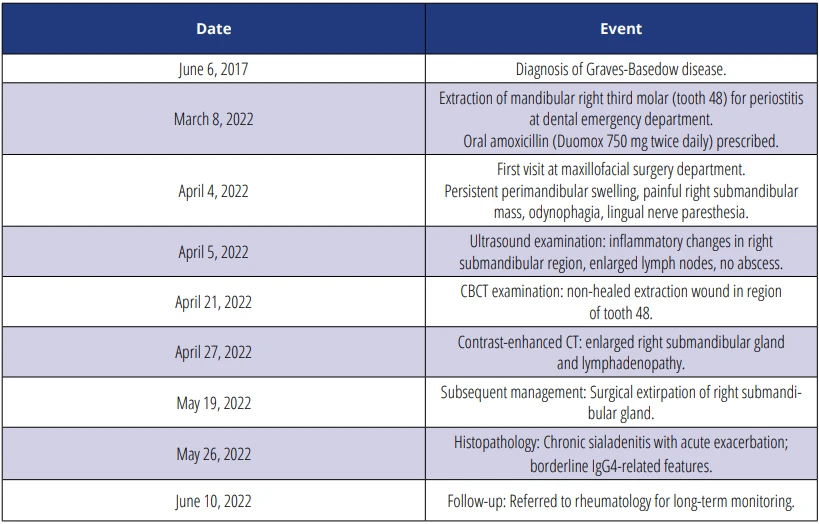
The patient was referred to our department on April 4, 2022, because of persistent symptoms following extraction of the mandibular right third molar (tooth 48, according to FDI classification), performed at a dental emergency department on March 8, 2022. The extraction of tooth 48 was undertaken to address periostitis, following which oral Duomox 750 mg (Amoxicillin trihydrate, Astellas Pharma s. r. o., Czech Republic) was prescribed twice daily. Post-extraction symptoms included persistent perimandibular swelling, a painful mass in the right submandibular region, odynophagia, and lingual nerve paresthesia.
Clinical assessment and preliminary diagnostic evaluation
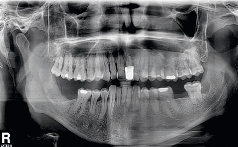
The initial examination revealed a firm, non-erythematous swelling in the right submandibular region with associated tenderness on palpation. The extraction site demonstrated incomplete healing, though without active exudation. Panoramic radiography revealed incomplete dentition with post-extraction changes in the site of extracted tooth 48. Initial diagnosis included residual inflammatory infiltrate in the right submandibular region and suspected early mandibular osteomyelitis.
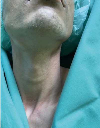
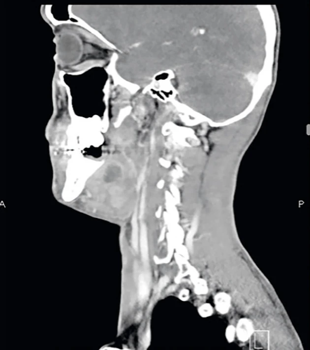
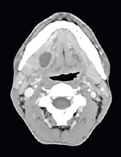
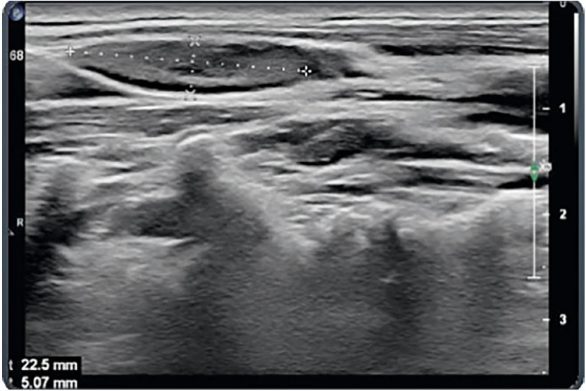
Ultrasound examination conducted on April 5, 2022, revealed inflammatory changes in the right submandibular region, with enlarged lymph nodes exhibiting normal oval morphology and no abscess formation. Subsequently, a CBCT scan performed on April 21, 2022, identified a non-healed extraction wound in the site of tooth 48. Due to persistent symptoms despite oral antibiotic treatment, a contrast enhanced CT scan was performed on April 27, 2022, which demonstrated enlargement of the right submandibular gland and lymphadenopathy (Fig. 1–5). Given the clinical diagnosis of a suspected submandibular gland tumor, surgical extirpation under general anesthesia was indicated.
Surgical intervention and histopathological findings
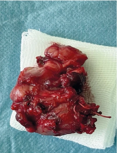
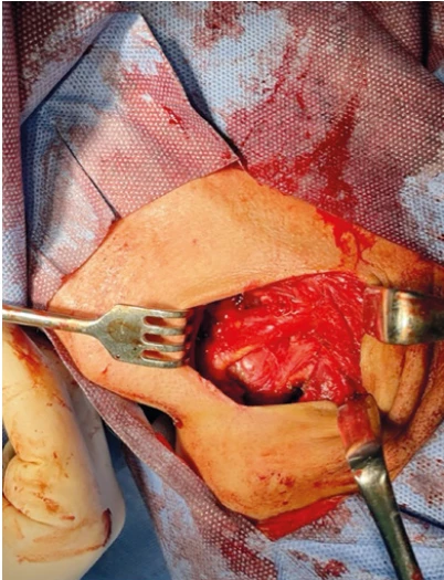
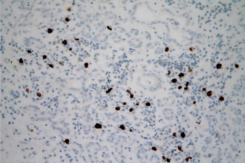
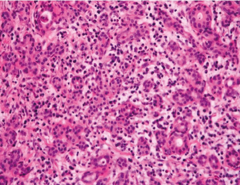
Surgical revision and right submandibular gland extirpation were indicated to exclude neoplastic growth, given the lack of clinical improvement. Histopathological examination revealed chronic sialadenitis with acute exacerbation, accompanied by fibrous septation and chronic lymphoplasmacytic inflammatory infiltration. No convincing obliterative phlebitis was identified. Immunohistochemical analysis demonstrated up to 30 IgG4-positive plasma cells per high-power field, with an IgG4/IgG tissue ratio of approximately 35–40%. Although the absolute number of IgG4-positive cells did not meet the diagnostic threshold for IgG4-related sialadenitis, the tissue ratio was borderline, supporting the need for further systemic evaluation (Fig. 6–9, Tab 1).
Post-operative course and follow-up
Post-operative recovery was uneventful with appropriate wound healing. Given the histopathological findings raising suspicion for IgG4-RD, the patient was referred to rheumatology for comprehensive evaluation and serum IgG4 level determination. From a surgical perspective, the patient's symptoms have been resolved, and no further follow-up visits at the maxillofacial surgery department were required. Based on the rheumatology department's assessment, the patient's condition requires long-term monitoring and observation, with no immediate treatment necessary, as the definitive classification remains unresolved, and long-term rheumatologic follow-up was recommended.
DISCUSSION
This case highlights the importance of comprehensive evaluation in cases of persistent submandibular swelling and the need to consider IgG4-RD in the differential diagnosis, particularly in patients with pre-existing autoimmune conditions. In the present case, differential diagnostic considerations included chronic inflammatory sialadenitis temporally associated with preceding odontogenic infection, reactive lymphoplasmacytic inflammation, and possible obstructive salivary gland disease. The patient’s pre-existing Graves-Basedow disease further complicated the clinical picture, as autoimmune comorbidity may coexist with salivary gland enlargement and alter serological markers.
These findings underline the necessity of integrating laboratory, histopathological, radiological, and clinical data when evaluating suspected IgG4-related disease.
IgG4-RD represents a complex immune-mediated systemic disorder characterized by distinctive tissue infiltration by IgG4-positive plasma cells accompanied by varying degrees of fibrosis. In physiological conditions, IgG4 comprises less than 5 percent of total immunoglobulin G. Although elevated serum IgG4 levels are characteristic of IgG4-RD, their precise pathogenic role remains incompletely understood. Recent studies have highlighted the role of plasmablast expansion, T helper 2 responses, regulatory T cells, and cytotoxic CD4+ T lymphocytes in disease pathogenesis [5, 6].
Current evidence suggests that IgG4 typically exhibits anti-inflammatory properties; however, in IgG4-RD, this regulatory mechanism appears to be dysregulated, resulting in persistent inflammation and progressive fibrosis [7, 8].
The clinical manifestations of IgG4-RD are remarkably diverse, with the capacity to affect multiple organ systems simultaneously or metachronously. The disease typically presents with tumor-like lesions that can involve numerous anatomical sites, including but not limited to the pancreas, biliary system, lacrimal glands, major salivary glands, pulmonary system, kidneys, aorta, meninges, and thyroid gland [9]. Diagnostic confirmation relies on comprehensive clinicopathological correlation incorporating histopathological findings, serological markers including serum IgG4 levels, imaging studies, and exclusion of alternative diagnoses. In tissue specimens, increased IgG4-positive plasma cells and an elevated IgG4/IgG ratio may support the diagnosis when interpreted in the appropriate clinical context [10].
IgG4-related sialadenitis is currently regarded within the broader spectrum of fibroinflammatory salivary gland disorders, and historical entities such as Küttner tumor and chronic sclerosing sialadenitis may overlap with this spectrum [11]. The pathological process is marked by a prominent lymphoplasmacytic tissue infiltrate and the presence of cytotoxic T cell populations. Notably, in cases involving the submandibular glands, the concurrent presence of sialolithiasis may complicate the diagnostic process [12].
Universally accepted diagnostic criteria are still lacking. However, the 2019 ACR/EULAR IgG4-RD criteria represent a significant milestone. This approach reflects the need to integrate clinical, serologic, radiologic, and pathologic data to classify patients confidently. These criteria have been approved by the European League Against Rheumatism Executive Committee and the American College of Rheumatology Board of Directors, signifying that the criteria set has been quantitatively validated using patient data and has undergone validation based on an independent data set [13].
Therapeutic management primarily centers on systemic glucocorticoid therapy, typically initiating with prednisolone 30–40 mg/day for 2–4 weeks, followed by a carefully monitored tapering schedule. Additional therapeutic options include B cell depletion therapy with rituximab and various immunosuppressive agents such as azathioprine, methotrexate, and cyclophosphamide, which may serve as steroid-sparing alternatives or adjunctive treatments [14, 15]. The therapeutic objectives encompass achieving and maintaining disease remission, preventing progressive fibrosis, and minimizing end-organ damage. Disease prognosis demonstrates considerable variability, largely dependent on the extent of organ involvement and individual response to therapeutic intervention.
This case illustrates the importance of considering IgG4-related disease in the differential diagnosis of persistent salivary gland enlargement and unexplained chronic inflammatory lesions of the head and neck region. However, overlap with infectious, obstructive, autoimmune, and neoplastic conditions may create substantial diagnostic uncertainty, requiring careful clinicopathological correlation.
The present case is of particular clinical interest because the initial presentation followed recent odontogenic inflammation, while the histopathological findings remained borderline rather than definitive for IgG4-related disease. This case highlights that diagnostically challenging enlargements of the salivary glands may exhibit partial IgG4-related features without fully meeting established diagnostic criteria, and such patients may require prolonged multidisciplinary follow-up.
CONCLUSIONS
This case demonstrates the diagnostic complexity of persistent submandibular gland enlargement with overlapping inflammatory and possible IgG4-related features. Although elevated serum IgG4 levels and partial histopathological findings raised suspicion for IgG4-related disease, established diagnostic criteria were not fully met.
The case emphasizes the importance of cautious clinicopathological interpretation, exclusion of alternative causes such as chronic inflammatory or reactive sialadenitis, and multidisciplinary collaboration between dental clinicians, surgeons, pathologists, and rheumatologists.
Patients with borderline findings may require long-term follow-up, as definitive classification may only become apparent over time.
Funding
Supported within the projects BBMRI-CZ LM2023033 and EF16_013/0001674 and from the Charles University Cooperatio programme, research area DENT.
Conflict of interest
The authors declare that they have no competing interests.
Declaration on the use of artificial
intelligence
Artificial intelligence (AI) was used for language revision and correction.
ChatGPT (OpenAI, San Francisco, California, USA).
Authors' contribution to the publication
Conceptualization: MD, WAA.
Clinica l data collection and case management: MD, WAA, LT.
Histopathological evaluation: JL.
Literature review: MD, WAA.
Writing – original draft preparation:
MD, WAA.
Writing – review and editing: JL, BP, LT.
Supervision: MD.
All authors have read and approved the final manuscript.
Patient consent
Written informed consent was obtained from the patient for publication of this case report and all accompanying clinical information and images.
MDDr. Masood Dehghan
Department of Dentistry
Faculty of Medicine in Hradec Králové, Charles University
University Hospital Hradec Králové
Sokolská 581
500 05 Hradec Králové
e-mail: dehghanm@lfhk.cuni.cz
Zdroje
1. Hamano H, Tanaka E, Ishizaka N, Kawa S. IgG4-related disease – a systemic disease that deserves attention regardless of one's subspecialty. Intern Med. 2018; 57(9): 1201–1207. doi: 10.2169/internalmedicine.9533-17
2. Wallace ZS, Katz G, Hernandez-Barco YG, Baker MC. Current and future advances in practice: IgG4-related disease. Rheumatol Adv Pract. 2024; 8(2): rkae020. doi: 10.1093/rap/rkae020
3. Umehara H, Okazaki K, Kawa S, Takahashi H, Goto H, Matsui S, Ishizaka N, Akamizu T, Sato Y, Kawano M; Research Program for Intractable Disease by the Ministry of Health, Labor and Welfare (MHLW) Japan. The 2020 revised comprehensive diagnostic (RCD) criteria for IgG4-RD. Mod Rheumatol. 2021; 31(3): 529–533. doi: 10.1080/14397595.2020.1859710
4. Della-Torre E, Lanzillotta M, Doglioni C. Immunology of IgG4-related disease. Clin Exp Immunol. 2015; 181(2): 191–206. doi: 10.1111/cei.12641
5. Liu C, Zhang P, Zhang W. Immunological mechanism of IgG4-related disease. J Transl Autoimmun. 2020; 3 : 100047. doi: 10.1016/j.jtauto.2020.100047
6. Lanzillotta M, Mancuso G, Della-Torre E. Advances in the diagnosis and management of IgG4 related disease. BMJ. 2020; 369: m1067. doi: 10.1136/bmj.m1067
7. Czarnywojtek A, Agaimy A, Pietrończyk K, Nixon IJ, Vander Poorten V, Mäkitie AA, Zafereo M, Florek E, Sawicka-Gutaj N, Ruchała M, Ferlito A. IgG4-related disease: an update on pathology and diagnostic criteria with a focus on salivary gland manifestations. Virchows Arch. 2024; 484(3): 381–399. doi: 10.1007/s00428-024-03757-0
8. Witte T, Schulze-Koops H.IgG4-related sialadenitis: IgG4 is helpful, but biopsies are still crucial. Arthritis Res Ther. 2015; 17 : 368. doi: 10.1186/s13075-015-0888-7
9. Beyer G, Schwaiger T, Lerch MM, Mayerle J. IgG4-related disease: a new kid on the block or an old aquaintance? United European Gastroenterol J. 2014; 2(3): 165–172. doi: 10.1177/2050640614532457
10. Kamisawa T, Okamoto A. IgG4-related sclerosing disease. World J Gastroenterol. 2008; 14(25): 3948–3955. doi: 10.3748/wjg.14.3948
11. Deshpande V, Zen Y, Chan JK, Yi EE, Sato Y, Yoshino T, Klöppel G, Heathcote JG, Khosroshahi A, Ferry JA, Aalberse RC, et al. Consensus statement on the pathology of IgG4-related disease. Mod Pathol. 2012; 25(9): 1181–1192. doi: 10.1038/modpathol.2012.72
12. Thompson LDR. IgG4-related sialadenitis. Ear Nose Throat J. 2021; 100(5_suppl): 531S–532S. doi: 10.1177/0145561319890153
13. Wallace ZS, Naden RP, Chari S, Choi H, Della-Torre E, Dicaire JF, Hart PA, Inoue D, Kawano M, Khosroshahi A, Kubota K,et al.; American College of Rheumatology/European League Against Rheumatism IgG4-Related Disease Classification Criteria Working Group. The 2019 American College of Rheumatology/European League Against Rheumatism Classification Criteria for IgG4-related disease. Arthritis Rheumatol. 2020; 72(1): 7–19. doi: 10.1002/art.41120
14. Hong X, Zhang YY, Li W, Liu YY, Wang Z, Chen Y, Gao Y, Sun ZP, Peng X, Su JZ, Cai ZG, Zhang L, He J, Ren LM, Yang HY, Li ZG, Yu GY. Treatment of immunoglobulin G4-related sialadenitis: outcomes of glucocorticoid therapy combined with steroid-sparing agents. Arthritis Res Ther. 2018; 20(1): 12. doi: 10.1186/s13075-017-1507-6
15. Nambiar S, Oliver TI. IgG4-related disease. [Updated 2023 Aug 8]. In: StatPearls [Internet]. Treasure Island (FL): StatPearls Publishing; 2026 Jan. Available from: https://www.ncbi.nlm.nih.gov/books/NBK499825/
Štítky
Chirurgia maxilofaciálna Ortodoncia StomatológiaČlánok vyšiel v časopise
Česká stomatologie / Praktické zubní lékařství
2026 Číslo 3
Najčítanejšie v tomto čísle
- DIAGNOSTIC COMPLEXITIES IN A SUSPECTED CASE
- OROFACIAL FEATURES OF MULTIPLE HAMARTOMA SYNDROME IN A CHILD
- 35 let České stomatologické komory
- Umělá inteligence, roboti a světové stomatologické špičky v Praze