
Dlaždicobuněčný karcinom kůže různého stupně – rozdíly v expresi CD10
Cutaneous Squamous Cell Carcinoma of Different Grades: Variation of the Expression of CD10
Although it has been claimed that invasive cutaneous squamous cell carcinoma (SCC) does not express CD10, very few examples have been investigated regarding this matter. The claim of our study is to investigate the expression in 20 cases of SCC of different grades of differentiation. For that, we randomly selected from our archives 5 SCC-keratoacanthomatous (KA)-type; 5 SCC non-KA, well-differentiated; 5 SCC, moderately differentiated; and 5 SCC, poorly differentiated. In all these cases, we performed an immunohistochemical study for CD10. We found expression of CD10 (either stromal or cytoplasmic) in all the cases. While stromal expression (either focal or diffuse) seemed to have no relation to the degree of differentiation of the tumor, cytoplasmic expression of the marker was not found in any of the cases of the poorly-differentiated group. We conclude that 1) stromal expression of CD10 is not lost in deeply invasive SCC, as previous literature has suggested; 2) lack of cytoplasmic expression of CD10 by cutaneous SCC can be considered as an additional prognosis factor to investigate in the future; 3) our results seem contradictory to the current view that has defended atypical fibroxanthoma as an anaplastic type of SCC, since the former expresses CD10 in virtually 100 % of the cases.
Keywords:
squamous cell carcinoma – CD10 – keratoacanthoma
Autoři:
A. Fernandez-Flores
Působiště autorů:
Service of Anatomic Pathology, Hospital El Bierzo, and the Service of Cellular Pathology, Clinica Ponferrada Spain
Vyšlo v časopise:
Čes.-slov. Patol., 44, 2008, No. 4, p. 100-102
Kategorie:
Původní práce
Souhrn
Přestože se udává, že dlaždicobuněčný karcinom kůže (DBK) neexprimuje CD10, bylo takto dosud vyšetřeno jen nemnoho případů. Účelem naší studie bylo vyšetřit tuto expresi u 20 případů DBK různého stupně diferenciace. Náhodně jsme proto z našeho archivu vybrali po 5 případech DBK – keratoakantomového (KA) typu a neKA typu dobře diferencovaného, středně diferencovaného a málo diferencovaného. Ve všech těchto případech jsme provedli imunohistochemickou reakci na CD10. Zjistili jsme expresi CD10 ve všech případech (buď stromální, či cytoplazmatickou). Zatímco stromální exprese (fokální či difuzní) zjevně neměla žádný vztah ke stupni diferenciace nádoru, cytoplazmatická exprese markeru nebyla zjištěna ani v jednom případě z málo diferencované skupiny. Uzavíráme, že 1. u hluboce invazivního DBK není ztracena exprese CD10; 2. ztráta cytoplazmatické exprese CD10 v DBK může být považována za další prognostický faktor; 3. naše výsledky se neshodují s literárními názory, které považují atypický fibroxantom za anaplastický typ DBK, protože atypický fibroxantom exprimuje CD10 v prakticky 100 % případů.
Klíčová slova:
dlaždicobuněčný karcinom – CD10 – keratoakantom
CD10 has been suggested as a tool in the differential diagnosis between superficial basal cell carcinoma (BCC) and (superficial) squamous cell carcinoma (SCC) (4). This is based on the finding of cytoplasmic expression by superficial BCC, in contrast to the superficial SCC that does not express the marker (4). Although it was claimed that this lack of expression seems to be preserved in invasive SCC (4), very few examples have been investigated regarding this matter (7), and some of them have shown stromal positivity for CD10 (4). On the other hand, others have found a relation between the expression of CD10 by oral SCC and its prognosis (3). The matter of the CD10 status in cutaneous SCC is even more important if one takes into account that SCC is one of the main differential diagnoses, when facing the possibility of an atypical fibroxanthoma (AF). The latter is, nevertheless, CD10 positive in nearly 100 % of the cases (1, 2, 5).
The aim of this study is to investigate the expression of CD10 by SCC of different grades of differentiation.
Material and methods
We selected 20 cases of SCC in four groups of 5 cases each: 1) SCC-keratoacanthomatous (KA)-type; 2) SCC non-KA, well-differentiated; 3) SCC, moderately differentiated; and 4) SCC, poorly differentiated. The criteria to include the cases in each group were evaluated as described in the last edition of skin tumors of the WHO book (6). The cases were randomly selected from our archives until each group was completed. In all the cases, the diagnosis and the grade of differentiation were confirmed before being included in the particular group.
We also performed an immunohistochemical study on sections from the paraffin-embedded block corresponding to each case, for CD10 (IgG1; Biocare Medical; clone 56C6; Ref. PM129AA; ready to use).
Results
Table 1 shows the cases which were included in the study, with clinical details, as well as with the immunohistochemical results. All cases showed certain pattern of immunostaining, although sometimes this was only stromal and focal. Cytoplasmic immunostaining was evidenced in the KA-type cases, as well as in the well differentiated (WD) and moderately differentiated (MD) cases (fig. 1), but no cytoplasmic expression was seen in the poorly differentiated SCCs.
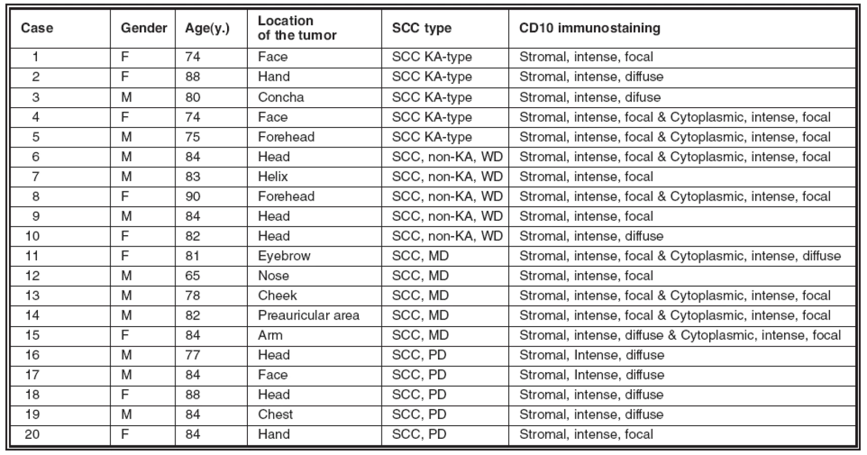

On the contrary, as the table also shows, different types of stromal staining were seen in all the cases (fig. 2).

Discussion
Some research groups have suggested that CD10 could have a diagnostic value in the differential diagnosis between superficial BCC and the (superficial) squamous cell carcinoma. This claim is based on the cytoplasmic expression of the marker by superficial BCC, opposite to what happens with superficial SCC, which does not express CD10 (4). It has been claimed that this lack of expression is preserved in deeply infiltrating cutaneous SCC, but very few examples have been investigated so far regarding this marker. Moreover, in some cases of infiltrating SCC, stromal positivity for CD10 can be found (4). On the other hand, a correlation between the expression of CD10 and the prognosis has been described in oral SCC (3).
Our results demonstrate that the stroma in invasive SCC expresses CD10 frequently, even if in a focal way only. Therefore, this is contrary to what has been suggested previously in the literature, that deeply infiltrating SCC does not express CD10 (4).
Our study also shows that the stromal CD10 expression seems not to be related to the grade of differentiation of the carcinoma. On the contrary, the cytoplasmic expression of CD10 was lost in all our cases of poorly differentiated SCC. This suggests the possibility of taking into account this lack of expression as a possible prognostic factor for the future. It should be, nevertheless, mentioned that other groups studying the expression of CD10 in cases of poorly differentiated SCC have found a patchy (weak to moderate) expression in up to 33 % of their cases (1).
The expression of CD10 by infiltrating SCC is also interesting in another context. Recently, it has been shown how atypical fibroxanthomas (AF) strongly express CD10 in nearly all cases (1, 2, 5). On the other hand, some authors have claimed that a strong relation between both entities exists, and that AF should be better interpreted as a variant of SCC (8). Nevertheless, the current findings would speak against such an interpretation, since CD10 expression seems to be lost when the good differentiation of SCC is also lost. Some other authors have also failed to find any connection between SCC and AF, when comparatively studying them (2), and our current results would support these latter studies.
Corresponding author:
Angel Fernandez-Flores, MD, PhD
S. Patología Celular, Clinica Ponferrada
Avenida Galicia 1
24400 Ponferrada
Spain
Telephone: (00 34) 987 42 37 32
Fax: (00 34) 987 42 91 02
e-mail: gpyauflowerlion@terra.es
Zdroje
1. Hultgren, T.L., DiMaio, D.J.: Immunohistochemical staining of CD10 in atypical fibroxanthoma. J. Cutan. Pathol., 34 : 415-419, 2007.
2. Mirza, B., Weedon, D.: Atypical fibroxanthoma: a clinicopathological study of 89 cases. Australas. J. Dermatol., 46 : 235-238, 2005.
3. Piatelli, A., Fioroni, M., Iezzi, G, et al.: CD10 expression in stromal cells of oral cavity squamous cell carcinoma: a clinic and pathologic correlation. Oral Dis., 12 : 301-304, 2006.
4. Wagoner, J., Keehn, C., Morgan, M.B.: CD-10 immunostaining differentiates superficial basal cell carcinoma from cutaneous squamous cell carcinoma. Am. J. Dermatopathol., 29 : 555-558, 2007.
5. Weedon, D., Williamson, R., Mirza, B.: CD10, a useful marker for atypical fibroxanthoma. Am. J. Dermatopathol., 27 : 181, 2005.
6. Weedon, D., Morgan, M.B., Gross, C., Nagore, E., Yu, L.L.: Squamous cell carcinoma. In: LeBoit, P.E., Burg, G., Weedon, D., Sarasin, A.: World Health Organization Classification of Tumours. Pathology and genetics of skin tumours. IARC Press: Lyon: 20-25, 2006.
7. Yada, K., Kashima, K., Daa, T., Gitano, S., Fujiwara, S., Yokoyama, S.: Expression of CD10 in basal cell carcinoma. Am. J. Dermatopathol., 26 : 463-471, 2004.
8. Zelger, B., Soyer, H.P.: Between Scylla and Charybdis: mythology in dermatopathology. Dermatopathol. Pract. Concept., 6 : 348-355, 2000.
Štítky
Patológia Súdne lekárstvo ToxikológiaČlánok vyšiel v časopise
Česko-slovenská patologie

2008 Číslo 4
Najčítanejšie v tomto čísle
- Dlaždicobuněčný karcinom kůže různého stupně – rozdíly v expresi CD10
- Co je nového v patologii štítné žlázy
- Heřman Šikl (1888–1955)
- Historky ze života Heřmana Šikla